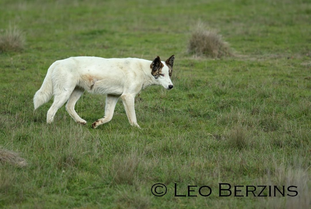
Wild dogs, including dingoes, feral domestic dogs and their hybrids cause significant economic loss to livestock production industries. Wild dogs also present a health hazard and can transfer diseases to humans and other animals. This research project investigated the potential transfer of the disease neosporosis from wild dogs to beef and dairy cattle in Australia.

Neosporosis is caused by the microscopic parasite Neospora caninum (N. caninum), which is a single-celled organism that can infect a number of animals including sheep, cattle, dogs and horses. Its life cycle includes several stages and the parasite may infect multiple animals over the course of its life. Neosporosis is a reproductive disease that causes abortions, low conception rates, decreased calving rates and milk production, and weaner and calf loss, and is estimated to cost the Australian beef and dairy industries in excess of AUD $110 million annually1.
Domestic dogs have been identified as definitive hosts of N. caninum2 – that is, the parasite begins and completes its life cycle inside the dog. Beef and dairy cattle act as intermediate hosts for N. caninum, where the parasite only passes through the cow’s body tissues. The N. caninum parasite is usually transmitted ‘vertically’ from the mother cow to its foetus (Figure 1). However, ‘horizontal’ transmission between domestic dogs and cattle also occurs when a dog eats N. caninum-infected tissue, such as placenta or aborted foetuses. The dog then sheds oocysts (ie N. caninum in egg form) in its faeces. Cattle then become infected by eating food and water contaminated by these oocysts.
Hypothetically, there is a second route of horizontal transmission: the proposed sylvatic route (Figure 1). This could involve wild dogs consuming N. caninum-infected tissue and either defecating on pasture and subsequently infecting wildlife, or consuming potentially-infected wildlife tissue. Although there is little evidence to support this type of transmission, the canine-bovine lifecycle may explain the association between many of the reported outbreaks of neosporosis in Australian cattle and their close proximity to bushland where wild canids are at high densities.
This project aimed to:
- Confirm the potential role of Australian dingoes in the transmission of N. caninum
- Determine the extent of N. caninum infection in wild dogs and remote community dogs.
Partners and management:
The case study was based on a PhD research project funded by the Invasive Animals CRC, the Dr William Richards Award and the NHP Graham Scholarship for sheep research at the University of Sydney. The Project Team included Jessica King (USyd), Prof Peter Windsor (USyd), Dr Jan Slapeta (USyd), Dr David Jenkins (Charles Sturt University), Dr Peter Fleming (NSW DPI), Dr Guy Ballard (NSW DPI) and Prof John Ellis (University of Technology Sydney).
Processes:
Aim 1 — Out of six newborn dairy bull calves that tested negative for N. caninum, two were infected with N. caninum at three weeks of age, and two at five weeks of age. The other two ‘control’ calves were left uninfected (Figure 2). The calves were euthanized at 10 or 12 weeks after infection, to be fed to dogs under experimental conditions. After confirming that all dogs were clear of N. caninum infection, four dingoes (one male and three females) at 12 weeks of age and four female domestic dogs between three and six months old ate an average of 1.3 kg of N. caninum-infected calf tissue. One of the dingoes and one of the domestic dogs were ‘control’ animals fed with meat from uninfected calves. An infected dingo (Dingo 3) was euthanized three days after it started shedding oocysts. Blood samples from all dingoes and dogs were collected for antibody and DNA analyses.
Aim 2 — Blood, serum and faecal samples from 75 wild dogs were collected from eight rural areas in New South Wales and Queensland between 2006 and 2009. Wild dogs were captured or shot by registered trappers and hunters for livestock protection. Samples from a total of 299 free-roaming community dogs were also collected from eight remote communities in Northern Territory, Western Australia, New South Wales and Queensland between 2000 and 2009. All samples from remote community dogs were collected as part of dog health programs within communities, and were provided opportunistically for this study.
It was not always possible to obtain all three samples from each dog.
Faecal samples were examined under a microscope for the presence of oocysts using a standard flotation technique, and faecal samples containing oocysts that were considered to be Neospora–like were processed for DNA extraction and analysis. Serum samples were screened for N. caninum antibodies, and DNA analysis was performed on the blood samples.
Results:
Dingoes and domestic dogs
Throughout the experiment, the calves showed no clinical signs of infection. The four infected calves tested positive to N. caninum by two to three weeks after infection and remained positive thereafter. The two calves that were not infected tested negative for N. caninum and no N. caninum DNA was detected in their blood. None of the dogs displayed any clinical signs of infection after eating the infected calf tissues. The presence of N. caninum DNA was confirmed by oocysts found in faeces collected from Dingo 3.
Wild dogs and remote community dogs
The presence of N. caninum DNA was also confirmed in faeces collected from two juvenile remote community dogs. Of the remote community dogs tested, significantly more adult dogs than juvenile dogs were infected with the parasite. It is likely that these dogs are becoming infected through their food. Although they are generally owned by a family, these dogs are not confined or tethered, and commonly have access to uncooked meat and other food waste, either provided by their owners or found through hunting or scavenging. A common dietary component for remote community dogs in the Northern Territory is meat from cattle, and to a lesser extent, macropods. These results suggest that remote community dogs may be involved in both the domestic horizontal transmission route of N. caninum in Australia (ie livestock to/from dog) and a sylvatic horizontal transmission route (dog to/from marsupial).
Recent experimental work has showed that the fat-tailed dunnart (Sminthopsis crassicaudata), a carnivorous marsupial widely distributed throughout the arid and semi-arid zones of Australia, was highly susceptible to neosporosis, thereby providing direct support for the existence of a sylvatic life cycle4. A wild dog from the Kosciuszko National Park in NSW has also tested positive to N. caninum. Antibodies detected in this adult wild dog suggest that exposure to N. caninum probably resulted from the consumption of natural prey within the area (eg marsupials, rabbits or rodents).
What worked:
The shedding of N. caninum oocysts from the intestinal tract of a dingo after eating infected meat provides direct evidence that Australian dingoes are definitive hosts of N. caninum.
Faeces were successfully screened for Neospora-like oocysts, and oocysts in faeces of two free-roaming dogs were identified. The shedding period is only short and shedding of N. caninum oocysts has only been documented in a few naturally-infected dogs worldwide.
What didn’t work:
No other dingo or domestic dog apart from Dingo 3 had detectable N. caninum DNA in their faeces before or after infection. However N. caninum DNA was detected in two of the infected dingoes and N. caninum antibodies were detected in two of the infected domestic dogs. This suggests that the numbers of parasites fed to the dogs in this study were possibly too low to cause detectable shedding of oocysts.
Dog 3 consumed tissues from the same two calves as Dingo 3, but oocysts were never detected in the faeces of Dog 3. This suggests that the pieces of tissue given to Dog 3 may not have contained the same amount of infectious material as the meat given to Dingo 3.
Conclusion:
This study provides evidence that dingoes are definitive hosts of N. caninum, and to our knowledge, is the first report of oocyst shedding by naturally-infected dogs (ie remote community dogs) in Australia. N. caninum is now found to be widespread throughout Australia and not just confined to NSW and Qld, where most of the previous studies on N. caninum have been focussed. The current study also confirms the potential routes for sylvatic transmission of N. caninum in Australia. However, farm dogs are likely to be as much to blame as wild dogs for the transmission of neosporosis to cattle. This is because farm and other domestic dogs have ready access to potentially infectious material through scavenging on dead cattle and afterbirth, or being fed raw tissue. The findings here also have implications for biodiversity conservation for native species, as the disease is potentially maintained by a sylvatic route where wild canids are the source of infection to their natural prey, such as marsupials.
Future research is required to determine the impact of removing dingoes from N. caninum-infected farms, as the relative significance of horizontal transmission compared to vertical transmission is unknown.
The hypothesis that Australian native wildlife may be carriers of N. caninum (ie the sylvatic route of horizontal transmission) also needs further investigation.
Further information:
- Hewitt L (2009). Major Economic Costs Associated With Wild Dogs in the Queensland Grazing Industry. Blueprint for the Bush and AgForce, Queensland, Australia.
- King JS, McAllan B, Spielman D, Lindsay SA, Hurková-Hofmannová L, Hartigan A, Al-Qassab SE, Ellis JT and Slapeta J (2011). Extensive production of Neospora caninum tissue cysts in a carnivorous marsupial succumbing to experimental neosporosis. The Veterinary Research 42:75.
- McAllister MM, Dubey JP, Lindsay DS, Jolley WR, Wills RA and McGuire AM (1998). Dogs are definitive hosts of Neospora caninum. International Journal for Parasitology 28:1473-1478.
- Reichel MP and Ellis JT (2009). Neospora caninum – how close are we to development of an efficacious vaccine that prevents abortion in cattle. International Journal for Parasitology 29:1173-1187.
See also:
- King JS, Slapeta J, Jenkins DJ, Al-Qassab SE, Ellis JT and Windsor PA (2010). Australian dingoes are definitive hosts of Neospora caninum. International Journal for Parasitology 40:945-950.
- King JS, Jenkins DJ, Ellis JT, Fleming P, Windsor PA and Slapeta J (2011). Implications of wild dog ecology on the sylvatic and domestic life cycle of Neospora caninum in Australia. The Veterinary Research 188: 24-33.
- King JS, Brown, GK, Jenkins, DJ, Ellis JT, Fleming PJS, Windsor PA and Slapeta J (2012). Oocysts and high seroprevalence of Neospora caninum in dogs living in remote Aboriginal communities and wild dogs in Australia. Veterinary Parasitology 187:85-92.
This PhD project by Dr. Jessica King was funded by the Invasive Animals Cooperative Research Centre (IA CRC) through the University of Sydney and was approved by the University of Sydney Animal Ethics Committee.
More information:
PestSmart toolkit for Wild dogs
Invasive Animals Ltd has taken care to validate the accuracy of the information at the date of publication [2014]. This information has been prepared with care but it is provided “as is”, without warranty of any kind, to the extent permitted by law.